Get to know the Anatomy of the Human Eye and How it Works
The eye is one of the body’s most important organs. You can see the green of the rice fields, the congestion on the road, and the rain on the windows because your eyes are functioning properly. Unfortunately, there are still many who do not know the anatomy of the eye and how to care for it properly. Come on, see the following reviews regarding eye images and their functions as well as tips for keeping eyes healthy.
Anatomy of the eye image and its functions
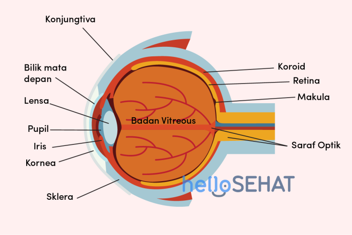
So that you can get to know more about the anatomy of the parts of the eye and their functions, consider the picture above and the explanation below.
1. Cornea
Cornea is a transparent dome-shaped network that forms the front or outermost part of the eye. The cornea functions as a window and a pathway for light to enter your eye.
Thanks to the cornea, your eye can control the entry of light rays so you can see words and pictures clearly. The cornea provides 65-75 percent of the focus power of your eye.
You also need to be careful to maintain the health of your cornea. In the cornea there are many nerve endings which make it very sensitive.
If not treated properly, the cornea is susceptible to bacterial or fungal infections such as keratitis. In addition, there is also the possibility of changes in the structure of the cornea, namely keratoconus.
2.The front eye chamber (anterior chamber)
The front eye chambers are sac-like jelly which is behind the cornea, in front of the lens (look at the image of your sense of sight above). The sac is also known as anterior chamber it is filled with liquid aqueous humor which helps carry nutrients to the eye tissue.
Fluid aqueous humor also at the same time serves as a counterweightpressure inside the eye. Eye health is also affected by the production process and flow of fluid in the front chambers of the eye. If there is interference, this can cause problems with the pressure inside the eye, such as glaucoma.
3. Sclera
The sclera is a hard white membrane-shaped part of the eye with fibrous tissue that covers the entire eyeball, except for the cornea. Inside are muscles attached to them to move the eye that is attached to the sclera.
Well, you also have to be careful because it does not rule out a problem with the sclera of the eye. One of the diseases associated with problematic sclera is scleritis, which is inflammation and swelling that occurs in the sclera.
4. Iris and pupil
The iris and pupil are part of the anatomy of the eye that are related to each other. The iris is a ring-shaped membrane that surrounds a small, darker sphere in the center.
Well, that little circle in the middle is called the pupil. The pupil is a muscle in the part of the eye that can close and open or shrink and enlarge.
Meanwhile, the iris functions to regulate the amount of light that enters the eye and adjusts to the pupil opening. When exposed to bright light, the iris closes (or narrows) and makes the pupil open smaller to limit the amount of light entering your eye.
Plus, it’s the iris that determines the color of your eyes. People with brown eyes have irises with lots of pigment. Meanwhile, blue-eyed people have irises with less pigment.
Iris and pupils are also not free from the possibility of disease. According to Mayo ClinicOne of the disorders that can occur is iritis, which is swelling and inflammation of the iris of your eye. Another name for iritis is uveitis.
5. Lens
The lens is the transparent, flexible part of the eye, which lies just behind the iris and pupil, after the cornea (see image of your sense of vision above).
The function of the lens is to help focus light and images on your retina. This lens provides 25-35 percent of the focus strength of your eye.
The eye lens has a flexible and elastic texture. Therefore, the shape can turn out to be curved and focus on the objects around it. For example, when you see people who are near you or from a distance.
The lens is also a common problem area of the eye. If someone has nearsightedness (myopia) or farsightedness (hypermetropy), this is caused by the incorrect position of the lens and cornea on the eyeball.
As we age, this important part of the anatomy of the eye can also lose its elasticity and the ability to focus on objects. This is commonly referred to as presbyopia or old eyes, namely visual disturbances that are experienced by many elderly people.
Another eye lens problem that often occurs as a result of aging is cataracts. This condition occurs when there is a spot or fog-like stain that covers part of the lens of the eye, so that the eye cannot see clearly.
6. Choroid and conjunctiva
The choroid is the part of the eye that is shaped like a dark brown membrane with many blood vessels in it. Its position is located between the sclera and retina.
This choroid serves to supply blood and nutrients to the retina and to all other structures in the anatomy of the eye.
Meanwhile, the conjunctiva is a thin layer of tissue that covers the entire front part of your eye, except for the cornea.
One of the eye disorders that can occur in the conjunctiva is conjunctivitis or pink eye. This condition is an inflammation and swelling of the lining of the conjunctiva, causing red and itchy eyes. Generally, this condition is triggered by a bacterial, viral, or allergen (allergen) infection.
7. Vitreous body
In contrast to liquids aqueous humor which is in front of the eye lens, vitreous humor located behind the eyepiece. Vitreous is a jelly-like substance that fills the inside of the back of the eye’s anatomy. Over time, the vitreous becomes thinner and can slip away from the back of the eye.
If your eye vision looks like there are white clouds floating around or flashing lights, see an ophthalmologist immediately. This is because a separate vitreous substance can cause a hole (a condition called a macular opening) to develop in the retina.
8. Retina and optic nerve
The retina is a tissue that is sensitive to light. This retina lines the inner surface of the eye’s anatomy. Cells in the retina can convert incoming light into electrical impulses. These electrical impulses are carried by the optic nerve (which resembles your television cable) to the brain, which in turn interprets them as the image or object the eye sees.
There are several eye problems related to the retina, which include:
- Retinal vein occlusion
- Cytomegalovirus retinitis
- A cut or tear in the retina
- Diabetic retinopathy
- Retinoblastoma
- Premature retinopathy
- Usher’s Syndrome
9. Macula
The macula is a small sensitive area in the center of the retina that provides central vision. In the macula, there is a fovea. The fovea is located at the center of the macula and its function is to provide the sharpest detailed vision in your eyes.
The macula is part of the anatomy of the eye with high levels of photoreceptor (light-receiving) cells that can detect light and send it to the brain. In other words, the macula has a big role so that you can see the various colors and details of an object very clearly.
Because its function is so crucial, damage to the macula can generally affect central vision or central vision.
One of the most common disorders in the macula is macular degeneration, which is an eye problem that usually occurs in people aged 50 years and over.
10. Eyelids
Even though it is located on the outermost part, the eyelid or palpebra is part of the anatomy of the eye with a function that is no less important than the other parts. The eyelids help maintain eye health by protecting your cornea from exposure to foreign objects, such as infections, injuries, and disease.
In addition, the eyelids also help to spread the tears evenly over the surface of the eye, especially if the eyelids are closed. This of course helps lubricate the eyes and prevent dry eye conditions.
However, you also have to be careful and keep your eyelids healthy. This is because the eyelids are prone to inflammation, infection, and other problems, such as:
Then, what do the eyes look like, aka the seeing process?
Each of the above eye anatomy parts work together so you can see clearly. However, what is the order in which they work?
First of all, light will enter through the cornea. After that, the cornea will regulate the entry of light into your eye.
Further light will pass through the pupil. Before that, the iris will be in charge of regulating how much light enters the pupil.
The light will then pass through the lens of the eye. The lens will work with the cornea to properly focus light on the retina of the eye.
When light hits the retina, the receptor cells convert the light into signals to be sent to the brain via the optic nerves. This way, your brain converts the signals into the pictures you are used to seeing.
Those are the 10 parts of eye anatomy along with their function and how they work that you must know. There are many ways you can do to maintain eye health, from adopting a healthy diet for your eyes, protecting your eyes from direct sunlight, to undergoing regular eye examinations to an eye specialist.