Heart Anatomy: Parts, Functions, and Diseases
The heart is an important organ that pumps blood in your body. If the heart and vessels have problems, it will certainly cause various heart diseases and cause symptoms. Worse, if the heart loses its function, death can occur. So, what is the anatomy of the heart and how does this organ work in your body? Let’s learn more in the following review.
Understand the anatomy of the heart and its functions
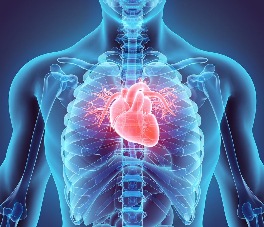
If depicted, the heart has a size slightly larger than your fist, which is about 200 to 425 grams. Your heart is located between the lungs in the center of the chest, behind and slightly to the left of the sternum (sternum).
For more details, let’s discuss one by one the anatomy of the heart with the image below.
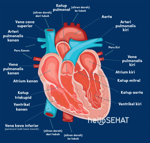
1. Pericardium
The heart is in a fluid-filled cavity called the pericardial cavity. The walls and lining of the pericardial cavity are called the pericardium. In the heart anatomy image above, the pericardium is in the middle.
The pericardium is a type of serous membrane that produces serous fluid to lubricate the heart during beating and prevent painful friction between the heart and surrounding organs.
This section also serves to support and hold the heart to stay in its position. The heart wall consists of three layers viz epicardium (outer layer), myocardium (middle layer), and endocardium (inner layer).
If you don’t keep your heart healthy, the pericardium can become inflamed and this is known as pericarditis. Meanwhile, if the endocardium and myocardium are inflamed, you will experience endocarditis or myocarditis.
2. Porch (atrium)
The porch or also known as the atrium is the upper part of the heart consisting of the right and left atrium. Right porch serves to receive dirty blood from the body carried by blood vessels.
While left foyer serves to receive clean blood from the lungs. The porch has thinner walls and is less muscular, because its job is only as a room for receiving blood. In the anatomical image above, the porch is on the right and left sides of the upper heart.
3. Chambers (ventricles)
Just like the atrium, the chambers or also called the ventricles are the lower part of the heart which consists of the right and the left. Right booth serves to pump dirty blood from the heart to the lungs. Meanwhile, left booth serves to pump clean blood from the heart to the rest of the body.
The walls of the chambers are much thicker and more muscular than the porch because they work harder to pump blood from both the heart to the lungs and to the rest of the body. In the anatomical image above, the ventricles are located on the right and left sides of the lower heart.
4. Valves
Look at the anatomy picture of the heart above, there are four valves that keep blood flowing in one direction, namely:
- Tricuspid valve, regulate blood flow between the right atrium and the right chamber.
- Pulmonary valve, regulates blood flow from the right ventricle to the pulmonary artery which carries blood to the lungs to pick up oxygen.
- Mitral valve, drains oxygen-rich blood from the lungs from the left atrium to the left ventricle.
- Aortic valve, opens the way for oxygen-rich blood to pass from the left ventricle to the aorta (the largest artery in the body).
In some people, the heart valves may not work properly, leading to valve heart disease.
5. Heart muscle
Cardiac muscle is a combination of striated and smooth muscle that is cylindrical in shape and has light and dark lines. When viewed closely under a microscope, this muscle has many cell nuclei located in the center.
The muscles in the heart are responsible for pumping blood throughout the body. The heart muscle is considered the strongest muscle because it is able to work continuously all the time without resting to pump blood. If this muscle stops working, the circulatory system will stop, resulting in death.
Well, in this heart muscle, you will get what is called the cardiac cycle, which is the sequence of events that occur when the heart beats. There are two phases of the heart cycle, namely:
- Systole, heart muscle tissue contracts to pump blood out of the ventricles.
- Diastole, the heart muscle relaxes when blood is filled in the heart
Blood pressure increases in the main arteries during ventricular systole and decreases during ventricular diastole. This results in 2 numbers related to blood pressure.
Systolic blood pressure is the higher number and the diastolic blood pressure is the lower number. For example, blood pressure 120/80 mmHg represents systolic pressure (120) and diastolic pressure (80). The heart muscle can weaken or have structural abnormalities, and this is known as cardiomyopathy.
6. Blood vessels
Look at the heart anatomy picture above, there are three main blood vessels in the heart, namely:
Artery
These heart blood vessels are rich in oxygen because blood functions to the left side of the heart muscle (left ventricle and atrium). Arteries have walls that are elastic enough to keep blood pressure consistent ..
The left main coronary artery then branches to form:
- Artery Left Anterior Descending (LAD), serves to provide blood to the top and left of the heart.
- Artery Left Circumflex (LCX), The left main artery branches around the heart muscle and provides blood to the outside and back of the heart.
The right coronary artery is responsible for supplying blood to the right ventricle, right atrium, SA (sinoatrial) and AV (atrioventricular). The right coronary artery branches into an artery Right Posterior Descending, and the right marginal artery. Together with the LAD, the right coronary artery helps supply blood to the lining of the heart.
Blood vessels in the heart can cause problems, such as coronary heart disease and atherosclerosis, both conditions signaling blockages in the heart vessels.
Veins
This one blood vessel carries oxygen-poor blood from all over the body to return to the heart. Compared to arteries, veins have thinner vessel walls.
Capillary
These blood vessels are in charge of connecting the smallest arteries with the smallest veins. The walls are so thin that they allow blood vessels to exchange compounds with the surrounding tissue, such as carbon dioxide, water, oxygen, waste, and nutrients.
How is the mechanism or how the heart organ works?
After understanding the anatomy of the heart and the functions of each of its parts, you can move on to discussing how the heart works.
The mechanism of action of the heart is related to the flow of blood in the body. In short, the circulation of blood that is pumped by the heart is from the body to the heart, then to the lungs back to the heart and returned to the rest of the body.
On the left side of the heart (note the anatomy of the heart above), low oxygen blood enters the heart through the two inferior and superior veins and into the right atrium. The atria will contract, the blood will flow to the right ventricle through the open tricuspid valve.
Once the ventricles are full, the tricuspid valve closes to prevent blood from flowing backward into the atria. At that instant, the ventricles will contract and blood leaves the heart through the pulmonary valve, into the pulmonary artery and into the lungs. Then, the blood will return to be rich in oxygen.
This oxygen-rich blood will be pumped and flow through the right side of the heart. Blood will pass through the pulmonary veins to the left atrium. The atria will contract and flow blood to the left ventricle through the open mitral valve.
When the ventricles are full, they close to prevent blood from flowing back up into the atria. As the ventricles contract, blood leaves the heart from the aortic valve, into the aorta, and circulates throughout the body.
Of course, you have to take care of this vital heart function. The goal, so that you avoid various heart diseases in the future. You can help keep your heart healthy by checking your heart rate at Hello Sehat.
Hello Health Group and Hello Sehat do not provide medical advice, diagnosis or treatment. Please check our editorial policy page for more detailed information.