Human Skin Anatomy and Its Various Important Functions

The skin is the largest organ in the human body. If stretched out, the skin of an adult’s body was about two square meters – it could cover a door. The extent of our skin serves to protect every muscle, tissue and important organs in the body. The skin also functions to help regulate body temperature as well as a sense of touch. In addition, human skin also plays a role as a producer of vitamin D which is important for bone health. That is why we must always maintain healthy skin. However, do you really know and understand the anatomical structure of your own skin? Come on, see the following explanation.
What is the anatomy of the human skin like?
The thickness and color of the skin can differ from person to person, depending on many factors – including genetics, race, age, and gender. There are also some people who have more hairy skin than others.
Apart from all that, the skin basically consists of 3 main layers:
Epidermis
The epidermis is the first and outermost layer of skin, the only layer of skin that can be seen by the naked eye. The anatomy of the epidermal skin is largely formed by a layer of keratinocytes, which produce keratin.
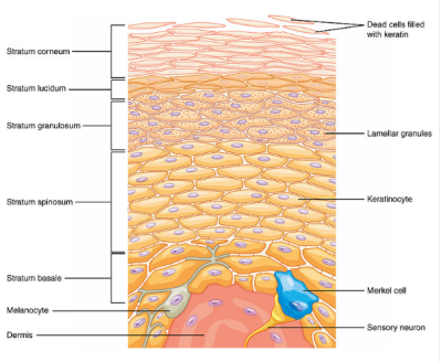
The epidermis itself is then further divided into 5 layers, namely:
- Basal stratum: the main keratinocyte production site
- Stratum spinosum: keratinocytes that are formed then bind to intercellular junctions called desmosomes
- Stratum granulosum: where skin cells produce fat and other molecules
- Stratum lucidum: functions to produce more keratin
- Stratum corneum: the top layer of the epidermis, which keeps producing keratin
Keratinocytes usually take between 30 and 40 days to travel from the stratum basale to the stratum corneum.
There are also 3 layers of non-keratinocyte cells that inhabit the epidermis, namely:
- Melanocytes: responsible for producing melanin (the pigment that gives skin color). The more melanin you produce, the darker your skin will be. Melanin production is influenced by your genetics.
- Langerhans cells: function as connective cells and skin’s defense system
- Merkel cells: function as a skin receptor
Dermis

The dermis is the second layer of skin after the epidermis. The dermis functions as a protector in the body. It is thicker in structure than the dermis, although it consists of only two layers – the superficial papillary layer and the reticular layer.
The reticular layer is much thicker than the papillary layer and contains clumps of collagen fibers.
Some of the cell structures that can be found in the dermis are:
- Fibroblasts: functions to produce collagen and elastin
- Mast cells: These cells contain granule histamine which comes from the immune system
- Skin appendages: the gathering place for hair follicles, sebaceous glands (oil glands), and sweat glands. Nail growth also starts here.
Subcutaneous (hypodermis)
The hypodermis layer is the innermost layer of the skin, which is also often referred to as the subcutaneous or subcutis layer. The subcutaneous layer contains the most fat to protect the body and help the body to adapt to outside temperatures. The hypodermis also acts as a skin binder to the muscles and various underlying tissues.
But don’t worry, the fat contained in this layer is not the same as bad visceral fat due to a bad lifestyle. The fat layer in the subcutaneous layer will always be under the skin. The amount can also vary for each individual depending on the composition of fat in the body.
Apart from containing fat, in this layer there are also many blood vessels.
Hello Health Group and Hello Sehat do not provide medical advice, diagnosis or treatment. Please check our editorial policy page for more detailed information.