Get to know the anatomy of the lungs from the start to its function

The lungs are the organs whose job is to process the incoming air and separate oxygen from carbon dioxide. This organ consists of two pairs, each of which has different characteristics. Intrigued by the function and what are the parts of the lungs? Come on, get to know more about the anatomy of this human lung.
What are the anatomy of the lungs and their functions?
Basically, the right and left lungs have different characteristics. The left lung of an adult weighs about 325-550 grams. Meanwhile, the right lung weighs about 375-600 grams.
Each lung is divided into several sections, called lobes, namely:
- The left lung consists of two lobes. The heart is in a groove (heart notch) located in the lower lobe.
- The right lung has three lobes. That is why the right lung is larger in size and weight than the left lung.
The lungs are separated by an area called the mediastinum. This area contains the heart, trachea, esophagus, and lymph nodes. The lungs are covered by a protective membrane known as the pleura and are separated from the abdominal cavity by a muscular diaphragm.
To know a more complete pulmonary anatomy, you can see the following picture.
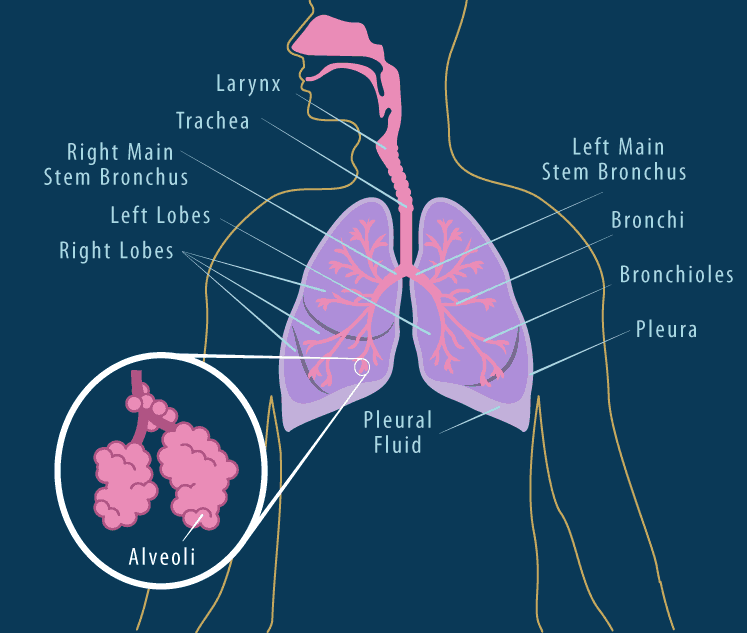
Summarized from Canadian Cancer Society, here is a complete explanation of the anatomy of the lungs:
1. Pleura
The first lung anatomy we will discuss is the pleura. The pleura is a thin, double-layered membrane that lines the lungs.
This layer secretes fluid (pleural fluid) which is called the serous fluid. Its function is to lubricate the inside of the lung cavity so as not to irritate the lungs when it expands and contracts whenbreathe.
The pleura consists of two layers, namely:
- Pleura (visceral), which is the lining next to the lungs
- The outer pleura (parietal), which is the layer that lines the chest wall
Meanwhile, the area that lies between the two layers is called the pleural cavity.
Some of the following types of diseases can arise when the pleura is problematic:
2.Bronchi (Bronchi)
The bronchi are the branches of the windpipe that lie after the throat (trachea) before the lungs. Bronchi are airways that ensure proper entry of air from the trachea to the alveoli.
Apart from being a route for the entry and exit of air, bronchi also function to prevent infection. This is because the bronchi are lined with various types of cells, including ciliated (hairy) and slimy cells. These cells later trap disease-carrying bacteria from entering the lungs.
If there is a problem with the bronchi, the following diseases can strike you:
3.Bronchioles (Bronchioles)
Each main bronchus divides or branches into smaller bronchi (having small glands and cartilage in their walls). These smaller bronchi eventually divide into even smaller tubes, called bronchioles.
Bronchioles are the smallest branches of the bronchi which do not have glands or cartilage. Bronchioles function to channel air from the bronchi to the alveoli.
In addition, bronchioles also function to control the amount of air that enters and leaves during the breathing process.
If this part of the lung is problematic, you can experience the following diseases:
4. Alveoli
This part of the anatomy of the lungs is the smallest group called the alveolar sac at the end of the bronchioles. Each alveoli is a concave-shaped cavity surrounded by many tiny capillaries.
The lungs produce a mixture of fat and protein called lung surfactants. This mixture of fat and protein coats the surface of the alveoli and makes it easier for them to expand and deflate with each breath.
Alveoli (alveoli) function as a place for exchange of oxygen and carbon dioxide. The alveoli then absorb oxygen from the air carried by the bronchioles and circulate it into the blood.
After that, carbon dioxide, which is a waste product from the body’s cells, flows from the blood to the alveoli to be exhaled. This gas exchange occurs through the very thin walls of the alveoli and capillaries.
If the alveolus is problematic, the following diseases can lurk you:
- Cardiogenic and non-cardiogenic pulmonary edema
- Pulmonary bleeding, usually due to vasculitis (eg Churge-Strauss)
- Pneumonia
- Alveolar protienosis and amyloidosis
- Bronchoalveolar carcinoma
- Alveolar microlithiasis
How do the lungs work?

Your lungs and respiratory system allow oxygen in the air to enter your body and let your body remove carbon dioxide in the air by blowing it out.
In breathing, your diaphragm moves up and your chest wall muscles relax. This causes the chest cavity to shrink and pushes air out of the respiratory system through the nose or mouth.
Your lungs and respiratory system will then perform the steps below:
- Each time you inhale, the air fills up most of the millions of alveoli
- Oxygen moves from the alveoli to the blood through the capillaries (small blood vessels) that line the walls of the alveoli
- Oxygen is taken up by the hemoglobin in red blood cells
- This oxygen-rich blood flows back to the heart, which pumps it through the arteries to the tissues, then around the body
- In the small capillaries of body tissues, oxygen from hemoglobin moves into the cells
- Carbon dioxide moves out of the cell into the capillaries
- Carbon dioxide rich blood returns to the heart via veins
- From the heart, this blood is pumped to the lungs, where carbon dioxide enters the alveoli to be exhaled outside the body.
Hello Health Group does not provide medical advice, diagnosis or treatment.
Chaudhry, R., & Bordoni, B. (2020). Anatomy, Thorax, Lungs. Statpearls Publishing. Retrieved from https://www.ncbi.nlm.nih.gov/books/NBK470197/
About your lungs – British Lung Foundation. (2015). Retrieved 18 August 2021, from https://www.blf.org.uk/support-for-you/how-your-lungs-work/about-the-lungs
The lungs – Canadian Cancer Society. (2020). Retrieved 18 August 2021, from https://www.cancer.ca/en/cancer-information/cancer-type/lung/lung-cancer/the-lungs/?region=on
(2020). Retrieved 18 August 2021, from https://gmch.gov.in/e-study/e%20lectures/Pathology/L%201.pdf
Anatomy of the Lung | SEER Training. (2020). Retrieved 18 August 2021, from https://training.seer.cancer.gov/lung/anatomy/
Pleural Diseases. (2016). Retrieved 25 August 2021, from https://www.bmc.org/pleural-diseases#:~:text=There%20are%20several%20types%20of,gas%20in%20the%20pleural%20cavity
Bronchial Disorders | Bronchiectasis | Bronchiolitis | MedlinePlus. (2020). Retrieved 25 August 2021, from https://medlineplus.gov/bronchialdisorders.html#:~:text=Bronchial%20disorders%20can%20make%20it,and%20become%20flabby%20and%20scarred
Bronchiolar Disorders | American Journal of Respiratory and Critical Care Medicine. (2020). American Journal Of Respiratory And Critical Care Medicine. Retrieved from https://www.atsjournals.org/doi/10.1164/rccm.200301-053SO