The Structure of the Human Ear: Pictures and Their Functions
Hearing is one of the supporting abilities in the human ear kom unicationone another. In addition, ears also function to maintain body balance. If your ears are experiencing interference, of course the activities you are doing will also experience obstacles. To find out more, see the following review of ear anatomy.
Understand the anatomy of the human ear
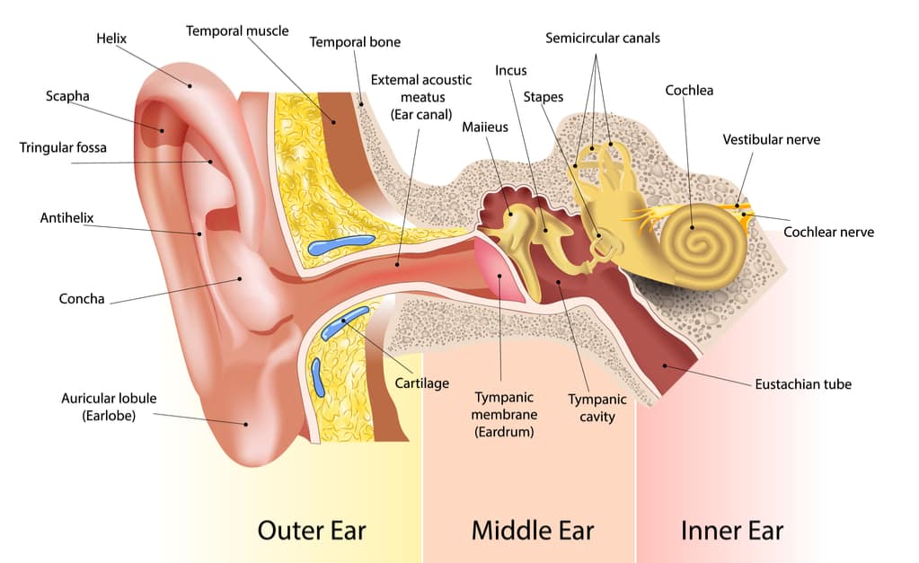
The human ear consists of three parts, namely the outer ear (outer ear), middle ear (middle ear), and finally the inner ear (inner ear). Consider the ear anatomy illustration based on the following three parts.
Outer ear (outer ear)
This ear structure is formed from the auricula (auricle) and the external auditory canal (the ear canal or ear canal). Auricula are formed by elastic cartilage that is tightly attached to sloping skin. This serves to capture sound and localize the sound. The auricula forms a hollow called the concha and its periphery is called the helix.
The structure of the earlobe consists of:
- Helical
- Spiral
- Antihelix
- Scaphoid fossa
- Triangle fossa
- Antihelix crura
- Antitragus
- Lobule
- Tragus
Ear canal (ear canal) formed by cartilage and temporal bone. It measures about 4 cm from the tragus to the tympanic membrane (tympanic membrane) which is also referred to as the eardrum and curves to form the letter S.
This arch is useful for preventing foreign bodies from reaching the tympanic membrane. There is a mandibular condyle in the front structure of the ear canal bone and a mastoid air cell at the end.
There are several sensory nerves in the outer ear, such as the auricular nerve, the occipital nerve, the ariculotemporal nerve, and the auricular branch of the phageal nerve (arnold nerve).
An ear disorder that you may face when the outer ear is problematic is otitis externa. This condition can also be referred to as swimmer’s ear.
Middle ear (middle ear)
The function of this part of the ear is to deliver the sound that the auricula has collected to the inner ear. This part of the ear extends from the cavity to the tympanic membrane, to an oval window consisting of the malleus, incus, and stapes bones and many intricate walls.
Tympanic membrane
The tympanic membrane is thin and semi-transparent, which separates the outer ear from the middle ear, which consists of pars flaccida and pars tensa. The malleus bone is firmly attached to the tympanic membrane in a hollow shape called umbo. The structure higher than umbo is called the flaccida pars and the rest is called the pars tensa.
There are three sensory nerves in the tympanic membrane, namely:
- Auriculotemporal nerve
- Arnold’s nerve
- Branch of the tympanic nerve
On the inner surface of the tympanic membrane there are chains of moving bones called ossicles, namely:
- Malleus (hammer)
- Incus (anvil)
- Stapes (stirrup)
These bone elements serve to conduct and amplify sound waves up to 10 times stronger than air to the inner ear.
Eustachian tube
The eustachian tube that connects the middle ear to the upstream of the esophagus and nose (nasopharynx). Its function is to equalize air pressure with the opening and closing movements. Important muscles found in the middle ear include the stapedius muscle and the tensor tympani tendon.
The horizontal part of the facial nerve crosses the tympanic cavity. Therefore, if there is paralysis of the facial nerves or muscles it will cause obstructed sound acuity and damage to the inner ear.
The following conditions may occur when your middle ear has a problem:
Inner ear (inner ear)
This ear structure is called the cavity labyrinth, which helps balance and transmits sound to the central nervous system. This cavity is formed from the osseous labyrinth, which is a series of temporal bones and a labyrinth of membranes (membrane sacs and channels). The membrane labyrinth also has components, namely:
Cochlea
Cochlea (cohclea) is an important organ in the inner ear which is shaped like a snail shell. It looks like a tube bent backward 2.5 circles with a cone at the end.
This section has three chambers, namely the vestibular scale, cochlear tract, and tympanic scale. In this cochlea, there is a core organ that functions to convert sound waves into nerve impulses.
Vestibular
The vestibular is the connecting part between the cochlea and the semicircular ducts. It consists of the sacula and utricula, which are hair cells that maintain a balance in the position of the head against the force of gravity when the body is at rest.
Semicircular
Semicircular is a semicircular canal of three different channels, namely horizontal semicircular canal, upper vertical semicircular canal, and rear vertical semicircular canal which contains ampules. This serves to determine awareness of the position of the head during rotational or rotating movements.
An ear disorder that you may face when your inner ear is problematic is labyrinthitis. In addition, sensorineural hearing loss also occurs when the inner ear, to be precise, the cochlear nerve is impaired.
How can you hear?

From ear anatomy, you have studied the structures that make up the ear, namely the outer ear, middle ear, and outer ear. The three parts of the ear become channels for external sound to enter and be translated into brain.
Reporting from Stanford Childrens, the process of hearing starts from the outer ear which picks up sound in the form of vibrations or waves around you. Then, the sound is lowered into the ear canal, causing pressure or a blow to the eardrum (tympanic membrane). When the eardrum vibrates, vibrations will be transmitted to the ossicles bone so that the vibration is amplified and sent to the inner ear.
Once the vibrations reach the inner ear, they are converted into electrical impulses and sent to the auditory nerve in the brain. The brain then translates these impulses as sound.
After knowing the ear anatomy, you will definitely understand that the ear is not only a hearing tool, but also maintains balance. This allows you to walk, jump, run without falling. When you feelear disorders You, immediately check your health to a doctor to get the right diagnosis and treatment.
Hello Health Group does not provide medical advice, diagnosis or treatment.